Lab 4: Sources of contamination and infection
Name of group member:
1. Chok Wen Xin (133291)
2. Leong Kah Yan (133310)
3. Yang Wen Huey(133376)
4.Joey Lew Hui Lin (133302)
Name of group member:
1. Chok Wen Xin (133291)
2. Leong Kah Yan (133310)
3. Yang Wen Huey(133376)
4.Joey Lew Hui Lin (133302)
Introduction
Airborne microorganisms are biological airborne contaminants also known as bioaerosols like bacteria, viruses or fungi as well as airborne toxins passed from one victim to the next through the air, without physical contact causing irritation. This usually happens when an infected subject sneezes, coughs, or just plain breathes. It is hard to prevent such a method of transmission.Airborne microorganisms are usually carried on dust particles, although some (fungal spores, for example) may be carried directly by air currents. Airborne microorganisms are a major cause of respiratory ailments such as allergies and pathogenic infections.Hence, it is important for microbiologists to be aware of the potential for contamination by airborne microorganisms. Carefully observation of simple precautions dramatically reduces the risk of contamination of the cultures.
Every human is colonized by billions of microorganisms. These microorganisms some of which are vital to our wellbeing, constitute our resident or normal microflora. Resident microorganisms are nourished by the chemicals and moisture excreted by the human bod. In moist areas such as the armpit, there may be one million bacteria per cm3; on the drier skin of the forearm, there may be 10000 bacteria per cm3.
Airborne microorganisms can be divided into two groups which are resident microorganism and transient microorganism. Resident microorganisms are either nonpathogenic or are prevented from infecting the body by an array of mechanical and chemical defences. Some resident microbes are, however, opportunistic pathogens which may cause infection if the body’s defences are breached-for example, if the skin is broken.
Transient or temporary skin flora refers to the microorganisms that transiently colonise the skin. This includes bacteria, fungi and viruses, which reach the hands, for example, by direct skin-to-skin contact or indirectly via objects.Transient microorganisms are picked up from our environment- for example from faecal contact or from soil- and usually fail to become permanent skin residents. One of the most important reasons for failure to gain permanence is that the established residents are better able to compete for nutrients. Since transients generally originate in other environments, they are poorly adapted to conditions on the skin and usually disappear within 24 hours of arrival.
Microorganisms in the upper respiratory tract are either normal residents or transients. As with the skin, the normal microflora largely consists of nonpathogens or opportunistic pathogens.
Large numbers of transients enter the upper respiratory tract as we breathe or eat. They may also come from our own hands or from improper sanitation during food preparation. Regardless of their origin, most transients are nonpathogenic and are quickly killed by various defences arrayed against them.
Objective
To determine the microorganisms in the air and from healthy human’s body
To determine the microorganisms morphology in culture medium agar.
Materials and reagents
Molten nutrient agar
Sterile water
Sterile petri dishes
Sterile clinical swab
Pipette and tips
Procedure
Air:
- The molten agar was poured into sterile petri dish and cooled.
- The lid was removed from the plate and leaved it resting on the side of the plate, facing down. (Never inverted the lid of the Petri dish). Leave the plates exposed for about 5 minutes.
- The lids were replaced and incubated at 37oC for 48 hours.
Hands:
- Hand was washed using sterile water. Do not use soap.
- 1 ml of wash water was transferred to the petri dish by using an automatic pipette.
- Molten nutrient agar was added to the Petri dish.
- The lids of the Petri dish were replaced and gently rotated the dish until the wash water was thoroughly mixed with the molten agar. Do not allowed the agar to contact the lid of the dish.
- After the agar had set, the dish was inverted and incubated at 37oC for 48 hours.
Ear:
- The molten agar was poured into sterile petri dish and cooled.
- Using extreme care,a sterile swab moistened with sterile isotonic solution were rubbed into the ear of the subject.
- Used the swab to inoculate the labeled plate.The inoculum was distributed as in the streak method.
- The inoculum was incubated at 37oC for 48 hours.
Normal breathing:
- The molten agar was poured into sterile petri dish and cooled.
- The lid was removed and the plate was hold about 15 cm from your mouth. Breathe normally but directly onto the plate for one minute. The lid is replaced.
- Incubated at 37oC for 48 hours.
Violent coughing:
- The molten agar was poured into sterile petri dish and cooled.
- The lid was removed and the plate was hold about 15 cm from your mouth. Coughed violently onto the agar. The lid was replaced.
- Incubated 37oC for 48 hours.
Result and observation:
1. Air
Commercial nutrient agar
|
Self-made nutrient agar
| |
Elevation
|
raised
|
raised
|
Margin
|
entire,undulate
|
Entire
|
Texture
|
moist
|
moist
|
Size
|
Small,large
|
small
|
Form
|
circular,irregular
|
Circular
|
Surface
|
Smooth,rough
|
smooth
|
Colour
|
Pale yellow and white
|
Pale yellow and white
|

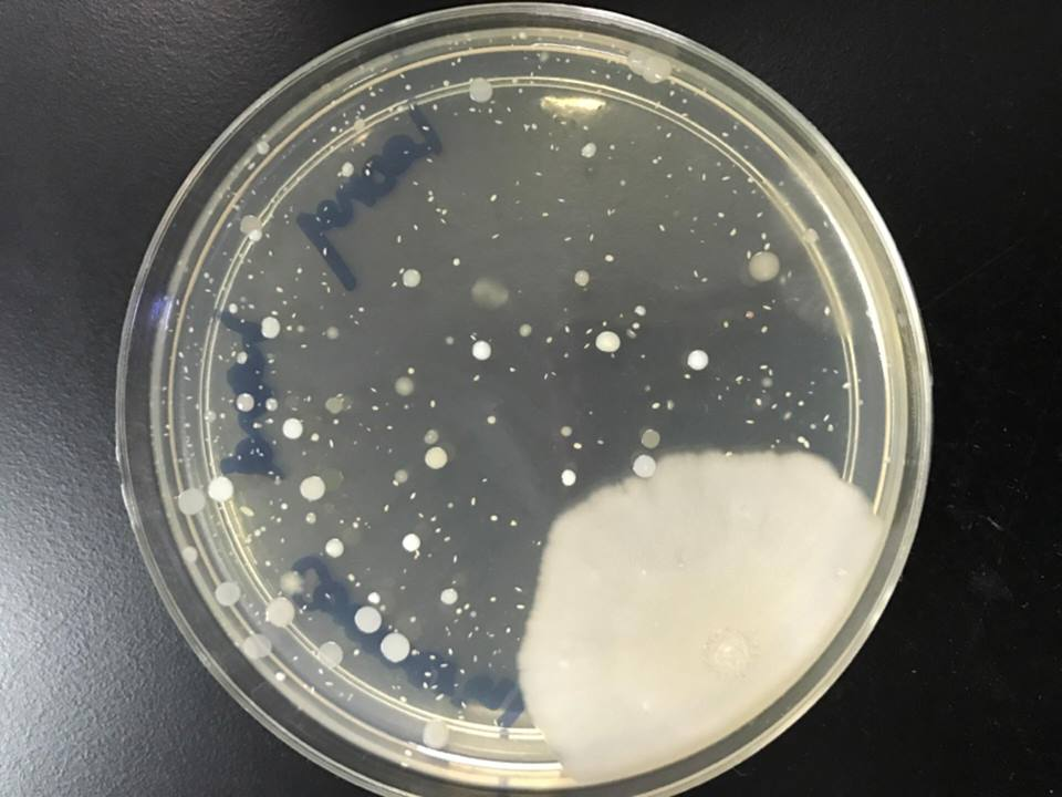
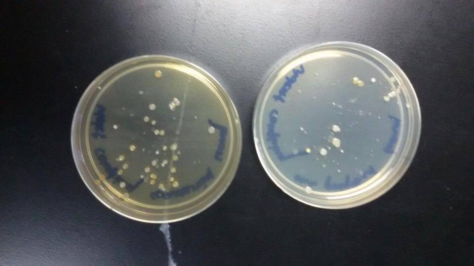
(Figure 1: sample from the air. Left: Self-made nutrient agar; Right:Commercial nutrient agar)
2. Hand
Commercial nutrient agar
|
Self-made nutrient agar
| |
Elevation
|
Raised, flat
|
raised
|
Margin
|
Entire,undulate
|
Entire,undulate
|
Texture
|
moist
|
moist
|
Size
|
Small,large
|
Small,large
|
Form
|
Circular,irregular
|
Circular,rhizoid,irregular
|
Surface
|
smooth,rough
|
Smooth,rough
|
Colour
|
white
|
White, yellow
|
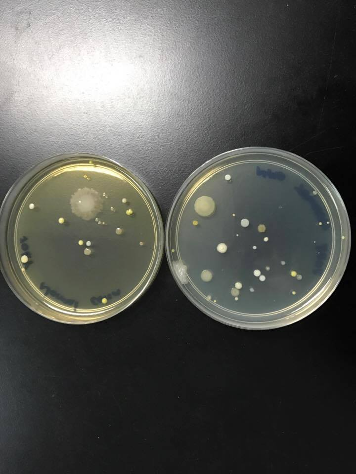
(Figure 2: sample from the hand. Left: Commercial nutrient agar; Right: Self-made nutrient agar)
3. Ear
Commercial nutrient agar
|
Self-made nutrient agar
| |
Elevation
|
raised
|
raised
|
Margin
|
undulate
|
undulate
|
Texture
|
moist
|
moist
|
Size
|
large
|
large
|
Form
|
irregular
|
irregular
|
Surface
|
rough
|
rough
|
Colour
|
white
|
white
|
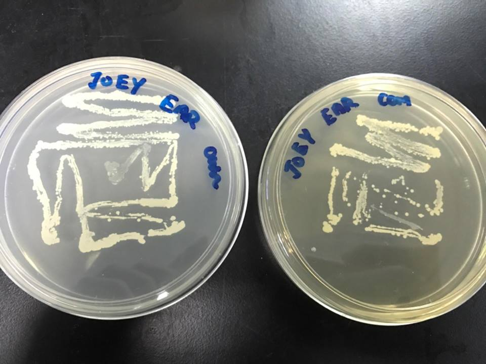
(Figure 3: sample from the ear. Left: Commercial nutrient agar; Right: Self-made nutrient agar)
4. Normal breathing
Commercial nutrient agar
|
Self-made nutrient agar
| |
Elevation
|
Flat,raised
|
raised,flat
|
Margin
|
entire
|
entire,undulate
|
Texture
|
moist
|
moist
|
Size
|
small,large
|
small,large
|
Form
|
circular,irregular
|
circular,irregular
|
Surface
|
smooth
|
smooth,rough
|
Colour
|
Yellow,white,transparent
|
white,yellow,transparent
|
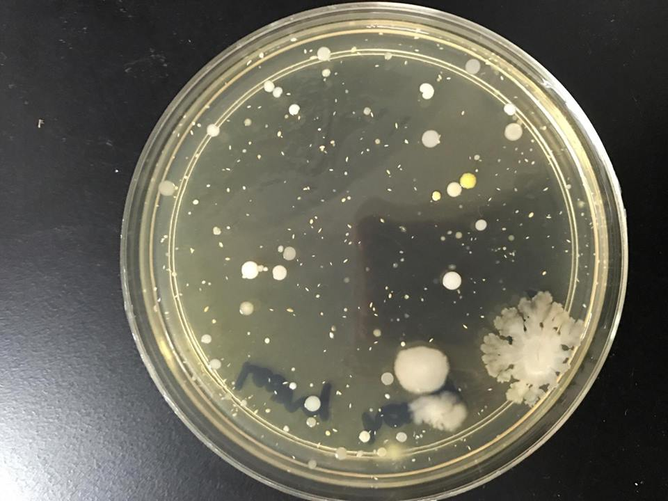
(Figure 4: sample from the normal breathing. Left: Commercial nutrient agar; Right: Self-made nutrient agar)
5. Violent coughing
Commercial nutrient agar
|
Self-made nutrient agar
| |
Elevation
|
Raised
|
raised
|
Margin
|
Entire
|
entire
|
Texture
|
moist
|
moist
|
Size
|
small
|
small
|
Form
|
Circular,irregular
|
circular,irregular
|
Surface
|
smooth
|
smooth
|
Colour
|
Pale yellow,white
|
Pale yellow,white
|
(Figure 5: sample from the violent coughing. Left: Commercial nutrient agar; Right: Self-made nutrient agar)
Discussion:
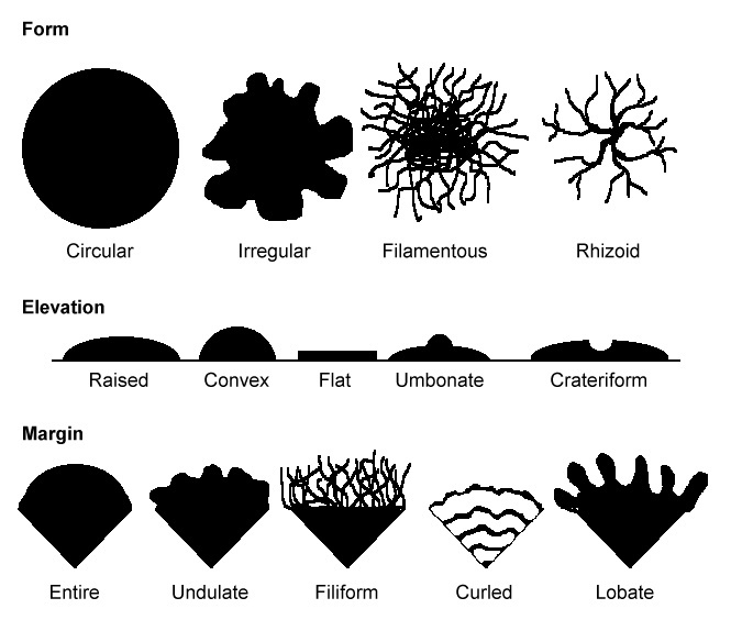
The differences between self made and commercial agar are compared and contrasted based on the morphology of the colonies.Different types of bacteria will produce colonies that have different appearances. Colony morphology is a method that scientists use to identify bacteria that grow on the agar according to their size, surface, texture, colour, form, elevation and margin.
From the sample of air contaminants , there were few colonies on the both media compared to the sample of other media that we have prepared.This is because the air in the laboratory is less contaminated. The common bacteria found in the air are Bacillus, Staphylococci and Clostridium. There are 2 type of colonies in self made agar and 3 type of colonies in commercial agar. On self made agar, the first colony are small, circular and white with raised elevation and smooth surface. The second colony are small, circular and pale yellow with raised elevation, moist texture and smooth surface. On commercial agar, the first colony are small ,circular and pale yellow with raised elevation, smooth surface and moist texture. The second colony are large, white and irregular with rough suface and undulate margin. The third colony are small, circular and white with raised elevation and smooth surface. Genally the colonies in both self made agar and commercial agar are distributed uniformly and there is no much differience between the colonies in self made agar and commercial agar.
From the sample of hand contaminants, we can found that there were many colonies formed. Our hand carries a lot of microbes because we usually come in contact with many contaminated things with our bare hand.There are different bacteria located on our hand some are good where else some are bad. There are some main pathogenic bacteria that can be found on our hand which is Haemophilus, Staphylococcus and Corynebacteria. There are 4 type of colonies in self made agar and 2 type of colonies in commercial agar. On self made agar, the first colony are small, circular and yellow with raised elevation and smooth surface. The second colony are small, circular and white with entire margin and smooth surface. The third colony are large, rhizoid and white with rough surface and undulate margin. The fourth colony are large, white and irregular with raised elevation and moist texture. On commercial agar, the first colony are small ,circular and white with raised elevation and moist texture. The second colony are large, white and irregular with rough suface and undulate margin. There is no much differience between the colonies in self made agar and commercial agar except the self made agar has rhizoid colony.
From the sample of ear contaminants, the colonies were formed along the line that we streaked. The common bacteia that can be found in our ear are Staphylococcus,Turicillotitidis and Alloiococcusotitis. The most common fungal microbe found in ear is Candida albicans.There are one type of colonies in both self made agar and commercial agar. The colony is large, irregular and white with undulate margin ,moist texture and rough surface. So, there is no different between self made agar and commercial agar.
From normal breathing contaminants, there are a some small of colonies found. These bacteria formed are non-pathogenic but some of them may induce illness. Bacteria that non-pathogenic are Streptococcus, Neisseria and Micrococcus. Bacteria that pathogenic are Staphylococcus aureus, Corynebacterium diphtheriae and Haemophilus. There are 3 type of colonies in commercial agar and 4 type of colonies in self made agar. There are three type of colonies are the same in both agar .The first same colony are small, white and circular with flat elevation and entire margin. The second same colony are small , yellow and circular with raised elevation and entire margin. The third same colony are transparent, irregular and smooth with entire margin and raised elevation. There is one colony in self made agar which the commercial agar do not have .The colony are transparent, irregular and rough with undulate margin and moist texture. So, there is no much different between self made agar and commercial agar.
From violent coughing contaminants,there are a lot of small colonies were found. This is because coughing can force air in our mouth that contain numerous of bacteria out of our mouth. So there more colonies can be found on the agar of violent coughing than the normal breathing. There are two type of colonies in both agar. The first colony is pale yellow , circular and smooth with raised elevation and entire margin. The second colony is white, irregular and smooth with raised elevation and entire margin. The amount of microorganism in commercial agar is greater than self made agar because the sample is taken from the same person and most of the microorganism entered the first plate which is commercial agar.
Conclusion:
From the experiment, we can found that there are not significant differences between the colonies formed in commercial nutrient agar and self-made nutrient agar. As long as the condition are favourable, the bacteria can grow well . We also can concluded that ear and hand have more contaminants compared to the others.
Reference: